Medicofit
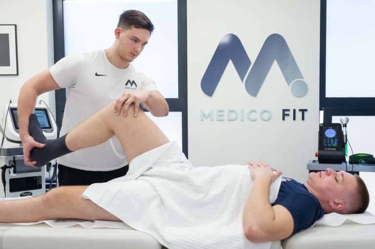
Sharing opinions, knowledge and experiences makes us stronger and better! We have prepared an overview of some successful physiotherapy and rehabilitation processes we have carried out, describing to you the process and the results in detail. If you find yourself in this description, don't hesitate to contact us today and join the crowd of satisfied customers!
TOP TENNIS PLAYER, LATERAL MENISCUS TEAR (BUCKET-HANDLE)
We considered the case of a 17-year-old tennis player who visited us 5 months after suturing a bucket-handle rupture of the lateral meniscus. The postoperative course was somewhat difficult, as the patient had to undergo four knee punctures due to extensive swelling and did not undergo rehabilitation at a spa due to school commitments.
At the time of inspection, the surgical scars were well healed and the knee was palpatory unremarkable. Mobility was full and pain-free, with only a tightening sensation on maximal flexion. All provocation tests for the medial and lateral meniscus were negative. Maximum isometric strength testing revealed a deficit of approximately 29% of the posterior thigh muscles and 18% of the anterior thigh muscles of the right leg, which is the most likely cause of the patient’s symptoms and non-sigmoid condition.
During the first month of treatment, we performed anterior and posterior thigh musculature strengthening uni- and bilaterally, both in open and closed kinetic chain. Due to the patient’s relatively fragile structure, the focus of the first phase of treatment was to increase overall strength and skeletal muscle mass. Over the course of ten treatments, he gained 1.5 kg of skeletal muscle mass thanks to graded strength training. Over the next 10 sessions, a transition to plyometric exercises of jumping, running and change of direction was followed, culminating in a Medicofit test battery.
FOOTBALL PLAYER, REHABILITATION AFTER ANTERIOR CRUCIATE LIGAMENT RECONSTRUCTION
A young football player was admitted after incomplete rehabilitation following anterior cruciate ligament reconstruction with associated medial meniscus suturing. During the examination, we noticed a 25-degree reduction in flexion mobility and a significant deficit in isometric strength of the anterior and posterior thigh muscles.
During the first 4 weeks, the focus was on strengthening the anterior and posterior thigh muscles and the ankle, knee and core stabilizers. After 4 weeks, a progression of jumping and running exercises was gradually introduced, followed by the last exercise of changing direction.
The rehabilitation process was extended by about 4 weeks from the planned 4 months, as the sutured meniscus was quite sensitive to eccentric-concentric loads, which had to be carefully dosed. On one occasion, the medial meniscus reacted to the load and on one occasion, mild inflammation of the pes anserinus occurred. In both cases of acute inflammation, laser and Tecar therapy were applied and the exercise process was adapted, which was never interrupted despite the reaction. Also, exercise rehabilitation was supplemented with HiTop high-frequency electrical stimulation throughout the treatment to accelerate the healing process.
After 5 months, the Medicofit test battery was successfully implemented and the return to training was completed during the preparation period for the 2022/23 season. The cooperation with the Medicofit clinic continues as part of the fitness preparation for the next season in the 2nd SNL.
BRAZILIAN JIU-JITSU ATHLETE, KNEE INJURY
We treated pain after a right knee injury. The gentleman had a right knee injury approximately 3 weeks prior to the Brazilian jiu-jitsu examination. The mechanism was contact, a hit to the outside of the knee. In addition to the injury, the gentleman mentioned intermittent pain on both distal attachments of the patellar ligament. The medical history also revealed a previous anterior cruciate ligament reconstruction of the right leg, which was an additional risk factor for the development of ipsi- and contralateral pathologies.
Given the non-acute phase of the injury and the mild symptomatology, we started with kinesiological therapy. In the first month, we focused on strengthening the extensor mechanism of the knee and strengthening the posterior thigh muscles in static and eccentric modes. Given the improvement in symptomatology and increased self-confidence, in the second month we made a transition to complex movement patterns and basic plyometric loads.
Although the patient’s injury was of a contact nature, due to a previous surgery, muscle asymmetries presented an increased level of risk for re-injury. We paid particular attention to training the posterior thigh muscles as muscle synergists of the anterior cruciate ligament.
After 10 weeks, the gentleman successfully corrected the muscle asymmetries, passed the internal lower limb test battery and returned to Brazilian jiu jitsu training.
POST-OPERATIVE KNEE REHABILITATION, ARTHROSCOPIC KNEE CARTILAGE RECONSTRUCTION WITH HYALOFAST MEMBRANE TECHNIQUE, BOXING SPORTS
A young boxer was admitted to our clinic 1 week after undergoing surgery. We found approximately 5 cm difference in thigh circumference between the operated leg and the non-operated leg. The mobility of the operated knee was painfully inhibited at approx. 60° of active and 70° of passive flexion, the extension was full but lagged behind the non-operated leg by approx. 5° of hyperextension. Quadriceps contraction was weak and unsustainable. On tensile testing, shortening of the posterior thigh muscles bilaterally was observed. On the day of the examination, more extensive joint oedema was present. The rehabilitation time to full return to competitive boxing training was estimated at 8 months.
The first phase of rehabilitation was performed in outpatient physiotherapy, improving mobility to a range of 130-140° of knee flexion. One of the goals of the acute post-operative rehabilitation was to reduce oedema, which decreased significantly and remained minimally present until the second phase of rehabilitation.
The knee loading protocol of the HYALOFAST method allows full knee loading after 6-8 postoperative weeks, when the patient also successfully abandoned crutches and started performing closed kinetic chain exercises. Initially, pain was experienced mainly with single-leg closed kinetic chain loading at 40-60° of knee flexion. During the first postoperative weeks, the thigh musculature was activated with HiTop electrical stimulation and BFR training (Blood flow restriction), and despite low mechanical loading, we managed to reduce muscle atrophy.
In the second phase of rehabilitation, the gentleman performed progressive closed kinetic chain loading, initially at low angles of knee flexion, gradually reaching 90° and 120° of flexion under full muscular load. Isolation exercises in the open kinetic chain were also applied aggressively, reaching 2/3 of the body weight in single-leg knee extension. We performed a Medicofit test battery for the knee and undertook teaching of running technique and transition to challenging plyometric exercises involving push-offs, jumping, turning and rapid changes of direction. The gentleman also received conditioning training throughout his rehabilitation to help him return to sport more effectively.
On the last day of rehabilitation, the patient tested positive on 5 Medicofit knee function tests and exceeded 95% symmetry between the legs on all of them, giving the green light to successfully complete the final ”return to sport” rehabilitation protocols.
BILATERAL ANTERIOR CRUCIATE LIGAMENT RECONSTRUCTION IN A YOUNG FEMALE BASKETBALL PLAYER
On the day of the diagnostic examination, two ACL reconstructions were diagnosed on both legs within a period of one year. The mechanism of injury of the right knee was contact and of the left knee non-contact. There was marked active and passive hyperextension of both knees, the ligamentous apparatus was firm. The rehabilitation goals were weight reduction, strength improvement, learning correct squatting and kicking techniques.
A 3 x weekly secondary rehabilitation programme was carried out, and given the complete absence of pain symptoms, treatment started in kinesiotherapy. We found weakness of the thigh muscles, uncontrolled knee stability and a more severe valgus knee position in squatting and push-off in jumping.
We started single leg strength training and improved the muscle strength of knee joint extension and flexion by more than 150% and achieved 95% symmetry of both legs. We progressively loaded the squatting and lifting patterns from the floor to successfully achieve the minimum standards for the plyometric part of the rehabilitation.
The patient successfully transitioned to running, rapid changes of direction and more advanced forms of plyometric exercise such as variations of single leg lunges and box jumps. The patient has successfully completed all 5 Medicofit Functional Tests for the knee joint.
The patient reduced body fat by 11% and gained 3 kg of muscle mass during rehabilitation.
NJURY TO THE POSTERIOR THIGH MUSCLE, BURNING PAIN ON THE BACK OF THE THIGH WHEN RUNNING OVER OBSTACLES.
A lady presented for a diagnostic examination for an injury to the posterior thigh muscle groups in the athletic discipline of hurdling. The lady did not undergo diagnostic imaging. Pain was present mainly in the lateral part of the middle third of the right posterior box. Since the injury, the patient has stopped running and only does strength and flexibility training and skipping in the pool.
On inspection, the patient was palpatorily painful in the lateral region of the middle third of the posterior lodge, corresponding to the biceps femoris. The mobility of the posterior thigh muscles was comparable to the uninjured side in the SLR test. Knee flexion and hip extension muscle strength tests showed pain.
The patient was assessed as having a posterior thigh muscle injury, specifically the middle third of the biceps femoris caput longum. To promote healing, we performed TECAR and LASER therapy and applied isometric strength exercises at different angles through knee joint flexion and hip joint extension. We performed a total of 6 weeks of rehabilitation using the 4+2 system. To the 4-week programme of progressive loading of the posterior gluteal muscle groups with strength exercises, we added a 2-week programme of sports rehabilitation with plyometric exercises and return-to-sport test protocols for the posterior gluteal muscles.
The rehabilitation programme was completed with higher intensity athletic exercises, and the patient was enrolled in the Medicofit physical training programme after rehabilitation.
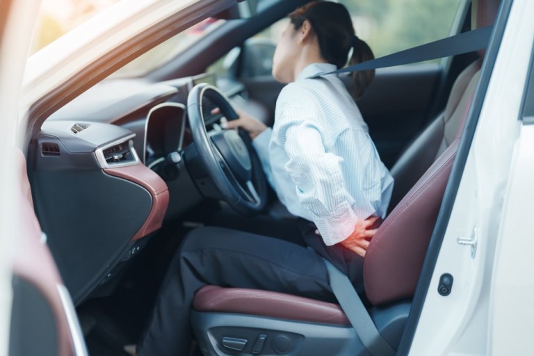
CHRONIC PAIN IN THE LUMBAR REGION AND BOTH ANKLES
The lumbar pain was mainly after prolonged periods of standing in place, occasionally with other movements. The patient had undergone an MRI scan in the past, which showed degenerative changes, mainly at levels L4 to S1. He had already tried manual therapy to manage the pain, which was unsuccessful until the visit to our clinic. The ankle pain was mainly present when weight-bearing while standing or lying on his back. MRI scanning showed posterior impingement syndrome with os trigonum. Previous treatment with FTH and infiltration blockade was unsuccessful.
We found mild extensor spinal intolerance and also that the sequence of hip and back extensor muscle activation during active hip extension was inappropriate in the patient. The inspection also revealed uneven development of the extensor musculature of the spine, in particular the caudal part of the extensors was significantly underdeveloped compared to the other parts.
A progressive therapeutic exercise programme was implemented, strengthening the core musculature through anti-rotation exercises, exercises to control pelvic positions and anti-extension exercises. We have gradually incorporated different variations of the plank exercise into the programme.
We strengthened the abductors of the hip joint, which were atrophied, in a targeted way and gained full tolerance to spinal extensor motion. We also taught a sequence of activation of the hip and back extensor muscles, followed by complex movement patterns such as squatting, dead lifting, overhead bending and carrying heavier loads.
The patient wanted to engage in higher intensity independent fitness training, so we performed an extended phase of posterior rehabilitation and achieved a 90 kg squat load for 10 repetitions without the onset of pain symptomatology.
ACHILLES TENDON TEAR IN BASKETBALL, POSTOPERATIVE REHABILITATION
On admission of the gentleman to the Medicofit clinic, thickening of the Achilles tendon and atrophy of the left calf musculature is observed, measured at 2.5 cm difference in circumference compared to the other leg. The ankle was restricted in the direction of dorsal flexion, approximately 5-10°. Muscle strength testing revealed a significant deficit in toe lifting strength, left (operated) leg – 11 lifts, right leg – 39 lifts. On the day of arrival, the patient was chronologically halfway through post-operative Achilles tendon rehabilitation according to our assessment, but was functionally delayed according to expectations and Achilles tendon rupture rehabilitation protocols.
We started treatment in kinesiotherapy with targeted exercises to correct the scapular musculature deficit. Dorsal flexion was improved over a 4-week period with active ankle mobilisation exercises, and the tibialis anterior muscle groups were strengthened to improve strength in the acquired range of motion of dorsal flexion. The gastrocnemius and soleus muscle groups were initially strengthened isometrically in different angles of plantar flexion with submaximal loading over a time span of up to 60 seconds. Submaximal eccentric exercises were gradually incorporated and applied in negative angles of plantar flexion, loading the Achilles tendon with tensile forces. Ankle stabilisation exercises were also performed in the medio-lateral direction.
The gentleman achieved 80% muscle strength symmetry after approx. 8 weeks, 90% symmetry after 12 weeks, a week later he passed the Medicofit functional test battery and started to switch to plyometric training.
Low impact plyometrics were performed on a coordinating ladder and with different variations of skipping using a stepper. After the initial plyometric session, there were no negative reactions of the Achilles tendon, so we made the transition to low-impact running on a treadmill and then on an artificial turf surface.
After 16 weeks, the patient started to perform ”high impact” plyometric training, which involved rapid eccentric-contralateral contractions of the Achilles tendon through bilateral and unilateral linked ankle jumps, where the goal was to minimise contact time with the ground.
After 20 weeks, the patient successfully completed the Medicofit plyometric test battery and achieved positive ”return to sport” protocols to resume playing basketball. During the rehabilitation process itself, conditioning and upper body strength training were also performed to prepare the patient for playing basketball.
SEVERE NECK PAIN, TINGLING IN THE ARM, KNEE PAIN, ACHILLES TENDON PAIN
A middle-aged gentleman was treated for severe pain deep in the neck and tingling in the left arm. The pain radiated to the inner side of the scapula and the tingling was mainly in the thumb, index and middle fingers of the left hand. The gentleman had already undergone an MRI scan prior to his visit to our clinic, which showed degenerative changes of the cervical spine between levels C5-C7. On examination, the gentleman also mentioned problems with his knee and Achilles tendon.
The gentleman first underwent manual therapy as part of his physiotherapy with us, as the levator scapulae and trapezius muscle groups had become over-toned as a result of the degenerative changes, and a certain degree of symptomatology from myofascial stiffness was noted. Instrumental therapy was applied minimally.
Physiotherapy included basic stabilization of the cervical spine together with corrective postural exercises, as there was also an increased curvature of the cervical spine (cervical lordosis). The gentleman continued to perform neurodynamic exercises to relax the nerve pathways, and after the tingling had resolved, the musculature of the cervical spine was strengthened by dynamic exercises and the rehabilitation was completed by teaching preventive techniques.
After the successful rehabilitation of the cervical spine problems, the gentleman continued with Medicofit personal training, where exercises to strengthen the thigh muscles and ankle muscle groups were performed, and a long-term exercise model for the management of cervical spine degeneration was established.
RIGHT SHOULDER AND UPPER ARM PAIN
Pain initially occurring in the elbow, which started after a prolonged period of working from home, was addressed. The pain moved to the upper arm and shoulder, with the greatest intensity at night and in the morning. The prescribed pharmacological painkillers did not alleviate the pain.
At the time of our inspection, there was intense pain in the triceps brachii, infraspinatus and trapezius muscles and throughout the shoulder joint. Active shoulder joint mobility was painfully inhibited in the anteflexion direction and in the abduction direction to approximately 80°. Passive mobility was slightly better, feasible up to 110° in the direction of elevation through abduction and through anteflexion. On performing the Spurling test, local pain in the cervical spine occurred, without radiculopathic signs.
In view of the symptoms present, we considered that the patient had calcification-related problems in the tendons of the infraspinatus muscle and problems associated with myofascial pain syndrome of the affected area.
We performed outpatient pain management with TECAR and LASER instrumentation. During the acute phase, only isometric exercises were performed and passive mobilization of the shoulder joint was performed. Shock waves were also applied but at a lower intensity. After 4 weeks, the patient started more dynamic exercises to improve rotator cuff strength, mainly through external rotation of the shoulder. We also performed stabilization exercises of the cervical spine and shoulder joint. Full mobility was restored after 6 weeks. After 12 weeks, the muscle function of the major muscle groups of the shoulder was suitable to perform the Medicofit battery test, which the lady passed positively.
INVERSION ANKLE SPRAIN WHILE PLAYING FOOTBALL
A young football player started treatment with us after an acute injury to his left ankle, which occurred while playing football. The gentleman was pre-treated at the emergency center, where he was diagnosed with an inversion ankle sprain. On our inspection, we found that the patient was most likely suffering from a high ankle sprain in the presence of anteromedial ankle compression syndrome.
The patient was treated with HiTop pain relief therapy as initially plantar flexion was severely inhibited as well as dorsiflexion (dorsal flexion, many articles are written under dorsiflexion). LASER and TECAR therapy were also performed to promote healing, with passive ankle flexion and active flexion within pain-free ranges of motion. The calf musculature was maintained by the application of electrical stimulation and isometric activation in non-painful intensity in angles that allowed activation.
Swelling was present at the time of our inspection and was significantly reduced in the first week. As the patient is an elite athlete, we performed 10 sessions per week of conditioning training, upper body strength training and thigh muscle strength training.
By the second week, we had already improved the range of motion tolerance of the dorsal and plantar flexors and gradually started progressive dynamic exercises to improve the strength of all functional directions of the ankle. Pain remained present in the dorsal most flexion. In the second half of the second week, we had already started ”low impact” plyometric training and the patient was back playing football at the end of the second week, but at a limited intensity.
At the end of the rehabilitation, in the third week, minimal pain symptomatology remained in the form of deep plantar flexion in the negative angle starting position, or eccentrically flexed dorsal flexion. The symptomatology is due to anteromedial ankle compression syndrome and the patient was prescribed regular preventive ankle exercises and has already started to play full competitive soccer.
LUMBAR SPINE PAIN, PRESENT FOR 20 YEARS
Chronic pain in the lumbar spine was described by the gentleman as a nagging sensation of stiffness and tightness. There have been more severe episodes of lumbar pain in the past, the most recent of which required him to take analgesics. There has been no ongoing leg pain.
On inspection of the lumbar spine, we observed a slightly flattened lumbar lordosis. On palpation, there was a marked increase in muscle tone of the spinal extensors, which were also palpatory pain-sensitive. Spinal mobility was severely restricted in the flexion direction, flexion was only possible up to mid-shin, lateral tilts were possible up to approx. 3 cm above the knee, and spinal extension was adequate. Sensitivity to right lateral rotation and anti flexion was observed when provocative patterns were tested. The mobility of both hip joints was slightly limited in the direction of internal rotation, the other directions were reasonably mobile. In the extended leg raise test, there was marked limitation of the mobility of the hamstrings and calf muscles. Cervical spine mobility was limited in all directions, without pain. Compression tests for the thoracic and cervical spine were negative.
The patient underwent manual release of the increased spinal extensor tone with concomitant core muscle strengthening exercises in kinesiotherapy as part of outpatient physiotherapy. Initial activation was performed in static postures aimed at strengthening spinal anti-rotation and anti-extension. We also performed mobility training for the hip joints and stabilization training for the cervical spine. The gentleman had poor control of his pelvic movement when performing basic strength exercises, so we included exercises to improve neuromuscular control of the sacro-lumbar spine. We strengthened the hip abductors with maximal isometric contractions and improved gluteal musculature endurance by 200%. On inspection, a strong gluteal activation was also observed.
The gentleman also started more dynamic strength exercises such as loaded squats and lifting loads off the ground. Given his long history of chronic pain, we advised him to join a long-term preventive exercise programme at the Medicofit clinic, which he attends twice a week under the supervision of a kinesiologist.
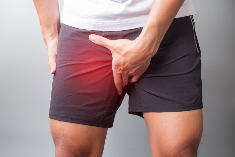
BILATERAL HIP PAIN.
A young female patient with chronic hip problems of 2 years’ duration was managed from the time of her diagnostic examination. MR arthrography showed an anterosuperior labral lesion of both hips. Pain was present even with a short distance walk and also when lying on her side. With initial anti-inflammatory therapy with TECAR and LASER, the conditions were achieved to start the application of isometric activation of the deep hip muscle groups.
As the pain was primarily in hip flexion and internal rotation, we started strengthening non-painful movements from the hip joint and gradually performed a progression of isometric specific exercises to gain tolerance to loading in flexion and internal rotation.
After gaining muscular endurance, pain in previously symptomatic directions decreased, so we made a progression to concentric forms of exercise in the hip joint and also started to gradually prepare the hips for longer distances of moderately fast walking.
Minor degenerative conditions of the hip joint are successfully treated by improving the muscular capacity of the deep hip musculature and building endurance in the strength of the key major muscle groups that perform hip flexion, extension, abduction and adduction.
WEAR AND TEAR OF THE RIGHT HIP JOINT, CHRONIC PAIN IN THE LUMBAR SPINE
The gentleman underwent preoperative preparation of the hip joint prior to total hip arthroplasty (joint replacement) in our clinic. On the day of arrival, the gentleman had medical records indicating degeneration of the intervertebral disc at the L4-L5 level, as well as spinal stenosis at the L3-L4 level and arthrosis of the facet joints. On consultation and examination, we conclude that the most likely cause of the pain is a combination of the overall pathological picture and suboptimal movement mechanics caused by a severely worn hip joint. On examination, we find an inability to maintain posterior pelvic tilt and reduced strength of the gluteal muscle group, particularly on the right side.
The gentleman in our clinic had lumbopelvic muscle strengthening, initially isometrically strengthening the spinal extension and core musculature, but with a range of movement that did not provoke symptomatology in the worn hip joint. We improved musculoskeletal control of pelvic movement and performed activation training for the deep hip musculature. The gentleman also undertook upper body physical preparation and healthy lower limbs, as one of the goals was a quick return to playing tennis after undergoing hip replacement.
The hip extension was highly symptomatic throughout the rehabilitation, so we performed it in a limited range of motion and gradually increased the tolerance to the load, but we did not perform more intensive exercise. BFR (blood flow restriction) training was also applied to the thigh musculature, which allows a hypertrophic effect with less mechanical stress on the joints.
The gentleman successfully rehabilitated his back pain, but until the hip replacement, we advised against sports activities and more intensive forms of exercise due to the suboptimal mechanics of movement of the entire hip and lumbar spine, which is aggravated by wear and tear of the joint (compensatory movements).
STRIATED CALF MUSCLE, EXTENSIVE RUPTURE OF M. SOLEUS DURING RUNNING
A gentleman underwent a diagnostic examination with us after re-injuring his right soleus. The gentleman had previously injured his right leg soleus muscle approximately 2 months ago while playing tennis. He underwent diagnostic ultrasound, which showed a large rupture of the soleus of the right leg in the middle third (80x20x30mm). At that time, a puncture of the haematoma was performed and compression was applied. The gentleman made good progress after the puncture. On follow-up examination, mild fibrosis of the injured part with a hematoma (approx. 3mm) was found. After follow-up examination, the gentleman resumed running training, which he did not have any problems with, but he did not do any targeted soleus strength training. One month after the follow-up examination, the gentleman recovered from the injury while running for a distance of approximately 3km.
On the day of the examination, palpatory findings showed a slightly more tender area in the medial head of the gastrocnemius, which is not related to the injury for which the gentleman presents for examination. Mobility of both ankles and knees was adequate in all directions. The length of the soleus muscle on the right shin was equal to that on the left, with a greater sensation of tightening at maximum extension than on the left leg. Isometric testing of soleus muscle strength revealed an imbalance between the left and right leg, which was strongly shifted in favor of the right injured leg. Single-leg toe raises in a squatting position were feasible without pain or loss of strength.
The patient’s problem was primarily due to a marked disproportion between the right and left leg. The re-injury was most likely due to a premature return to running training, without adequate prior strength training to allow sufficient adaptation of the healed tissue to higher loads.
The patient underwent progressive training to strengthen the calf musculature through the knee extensor and flexor. The patient performed isometric strength training m. Soleus and m. Gastrocnemius, and later we applied maximal concentric loads and forced eccentric loading. The gentleman also underwent a transition to plyometric training, learning running technique, and ”high impact” testing was performed using a Medicofit battery in the form of linked sonic box jumps with a rapid contact phase between jumps. The gentleman also performed an endurance plyometrics workout.
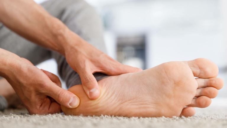
PAIN ON THE PLANTAR FASCIA AT THE CALCANEUS, HEEL SPUR
On the day of examination, the lady tells that the problems started in November 2020 and persist with relatively the same intensity until today. She underwent an X-ray and diagnostic ultrasound which showed both the presence of a calcaneal spine and thickening of the plantar fascia at the calcaneus. Until our visit, she had undergone a cycle of FTH, which had not resulted in any significant improvement. She has also had PRP applied, which has so far made the condition slightly worse. In her free time, the lady goes for long hikes in the mountains, and she also walks a lot at work, about 10,000 steps a day.
On inspection, the palpatory tender area of the plantar fascia process on the calcaneus is slightly more on the medial side. Toe mobility was comparable to the left foot. The right ankle was slightly restricted in dorsal flexion, approx. 5° deficit, and plantar flexion was comparable compared to the left ankle.
When testing ankle muscle strength, we found a significant deficit in plantar flexor endurance strength, with the patient performing 18 full toe raises on the left leg and only 12 on the right. When performing the Windlass mechanism, there was an increased stiffness of the right plantar fascia with the left.
The patient presented with plantar fasciopathy rather than classic plantar fasciitis, as indicated by the long-standing pain and the failure to treat the problem with conventional anti-inflammatory agents.
Outpatient physiotherapy included shock wave therapy and special exercises to strengthen the intrinsic muscles of the foot. Mobility deficits were corrected by the application of active mobility exercises and the plantar fascia was strengthened by prolonged isometric contractions. After each therapy, a high-energy pain-relieving LASER was applied to the plantar fascia and calcaneus. In the second phase of rehabilitation, we assessed and restored the biomechanical gait pattern and the correctness of foot positioning during gait.
After the end of the treatment, the lady joined the preventive programme of the Medicofit clinic, which includes general fitness training, exercises to improve joint functionality and unilateral and bilateral strength exercises.
POST-OPERATIVE REHABILITATION – SHOULDER SURGERY, BICEPS TENODESIS
A young football goalkeeper injured himself when parrying a ball and it hit his outstretched arm. He initially tried to repair the injury non-operatively, but rehabilitation was unsuccessful. The patient then underwent surgical repair of the SLAP lesion with biceps tendon tenodesis. The postoperative course was uneventful and the patient reported no pain on examination.
The arthroscopic wounds were healed on the day of the examination, without pain or tenderness to touch. There was atrophy of the deltoid muscle and periscapular musculature of the left shoulder. The mobility of the operated shoulder was good, almost perfect. The mobility deficits were mainly manifested in the end ranges of motion of elevation through anteflexion and elevation through abduction. Mild compensatory movements of the scapula were observed when performing the final elevation. The range of motion in external and internal rotation was almost equivalent to the non-operated shoulder, both in the prone position and in the 90° abduction position. The elbow and wrist were fully mobile in all directions.
The patient received physiotherapy 3 times a week in our clinic to restore full function of the operated shoulder joint, followed by kinesiotherapy with the same weekly regimen. The treatment consisted of intensive specialized therapeutic exercise, initially within post-operative limits and subsequently at full load. External rotation of the shoulder joint was performed in the scapular plane and in the adducted elbow-body position. Targeted strengthening of the m. Serratus anterior and by activating the lower and middle muscle fibers of m. Trapezius to reduce the present dyskinesia of the scapula. The patient underwent progressive strengthening of unilateral upper body strength patterns as part of kinesiotherapy, starting in the horizontal plane and gradually incorporating pure vertical strength training components.
As the patient was a professional athlete, we performed a ”high impact” return to sport test battery from the Medicofit clinic, which the patient passed with flying colors. During rehabilitation, we already included elementary ball exercises and progressively increased the shoulder load to catching and throwing heavier medicine balls. The patient started progressive integration into football training already during rehabilitation at the Medicofit clinic, and full competitive load was allowed after the return to sport test protocols were performed.
YOUNG FOOTBALL PLAYER, SURGERY ON THE MENISCUS
We admitted the young football player 5 days after a partial medial meniscectomy. During inspection (v slovenščini: Ob inspekciji. Tukaj bi mogoče raje napisala – During the examination?), an oedema on the right knee was discovered, measured at 2 cm difference over the mid-joint compared to the contralateral leg. Active knee extension was lagging by approximately 5°, passive knee extension was full. Active knee flexion was 105° and passive knee flexion was 110°. 20 cm above the kneecap, the circumference was 1.5 cm greater on the unoperated leg. The treatment time was estimated to be 8 weeks with a regimen of 5x weekly. As the football player is a high-level athlete, the treatment included intensive aerobic exercise, which was later escalated to a return-to-play phase of rehabilitation.
The first phase of rehabilitation was carried out in the context of outpatient and exercise physiotherapy. In 10 sessions, knee mobility improved from 105° of flexion to approximately 130°, and extension improved from -5° to 0°. The quadriceps circumference increased by approx. 2 cm. Knee edema has decreased. The patient started an introduction to strength training.
After 2 weeks, a transition to the kinesiological part of the treatment and progressive loading of the extensors and flexors of the knee followed. After demonstrating competence in lower limb strength and contralateral symmetry, we started a gradual progression of plyometric loading. The course of the injury itself was greatly facilitated due to good pre-existing muscle strength and general fitness. After 8 weeks, a positive Medicofit lower limb test battery was performed and a return to training was implemented.